
RESUMEN
Las metástasis espinales representan una importante carga sobre la calidad de vida en los pacientes afectados por una enfermedad oncológica activa, debido a la alta incidencia de síndromes dolorosos, deformidad espinal y deterioro neurológico. La cirugía juega un papel determinante a la hora de mejorar la calidad de vida mediante el control del dolor, el restablecimiento de la función neurológica y el mantenimiento de la estabilidad espinal, además de contribuir a la respuesta de la terapia médica. La cirugía mínimamente invasiva es una opción de tratamiento en determinados pacientes con alto riesgo quirúrgico, ya que tiene una baja tasa de complicaciones, de sangrado intraoperatorio, de estancia hospitalaria y ofrece resultados similares a la cirugía abierta.
Presentamos en esta revisión el papel de la cirugía mínimamente invasiva en esta enfermedad, y algunos casos tratados en nuestro centro hospitalario.
Continua en inglés
Introduction
In the United States, cancer diagnoses affect approximately 1.6 million people each year, with a mortality rate of around 50%. This disease places a significant burden on both patients and the healthcare system. Spinal metastatic involvement is a common complication of cancer, and its prevalence is increasing due to advancements in complementary treatments such as chemotherapy and hormone therapy, which have improved patient survival rates. After lung and liver involvement, the spine ranks as the third most frequent site for metastases, primarily due to prostate, lung, and breast cancers. Spinal metastases occur in 30% to 90% of cancer patients, but only about 10% of these patients experience symptoms related to spinal involvement, with roughly 50% requiring some form of treatment, and 5% to 10% needing surgical intervention.
Spinal Involvement
Tumors can disseminate through various mechanisms: hematogenous spread, contiguous invasion, or pathological seeding in the cerebrospinal fluid (CSF). Hematogenous dissemination is the most common method, taking advantage of the rich arterial blood supply in vertebral bodies to migrate tumor cells from their origin to the spine. Dissemination through Batson’s venous plexus is also possible due to extensive connections between this network and other venous drainage pathways. Regardless of the route, this type of dissemination often leads to multiple spinal lesions.
Clinical Presentation
Spinal metastases can manifest with varying symptoms, but pain is the most prevalent, affecting up to 95% of cases. This pain can be local, mechanical, or radiating. Motor dysfunction, occurring in up to 85% of patients, typically presents as weakness in one or more muscle groups and may result from myelopathy, radiculopathy, or a combination of both.
Treatment
The treatment of spinal metastases encompasses various modalities and medical specialties, with the choice depending on factors such as the patient’s clinical condition, life expectancy, primary tumor control, predominant symptoms, extent of spinal involvement, and more. Surgical intervention is generally considered for patients with a life expectancy of more than 3 to 6 months. It’s important to note that the primary goal of treatment for spinal metastases is palliative, aimed at ensuring patient safety and improving their quality of life.
Surgical Treatment
When surgical treatment is indicated, the objectives are to alleviate or eliminate pain, preserve or enhance neurological function, and maintain or restore spinal stability. Spinal instability due to neoplastic involvement is a surgical indication, regardless of the degree of epidural compression. The Spine Instability Neoplastic Score (SINS) helps evaluate the degree of instability, aiding surgeons in identifying at-risk patients.
Advances in Surgical Techniques
Minimally invasive surgery (MIS) has emerged as an alternative to traditional open surgery for treating spinal oncological diseases. MIS offers advantages such as reduced blood loss, shorter hospital stays, better pain management, and decreased postoperative opioid use. Comparisons between open surgery and MIS have shown similar outcomes in terms of neurological improvement and pain control. However, it’s crucial to emphasize that the mastery of MIS requires a significant learning curve.
Clinical Cases
Let’s delve deeper into three clinical cases that illustrate the application of minimally invasive surgery (MIS) in treating spinal metastases:
Clinical case 1
A 46-year-old woman with a history of breast cancer attended the emergency department for progressive loss of strength in the lower limbs until she was unable to ambulate at <24h (SOSG) from onset.
Physical examination: Lower limb (R/L): hip flexion 3-/3-, hip extension 5/5, knee extension 5/5, knee flexion 5/5, dorsal and plantar flexion 5/5, and sensory level D6.
An emergency MRI of the dorsal spine was performed, which showed multilevel involvement of the dorsal spine (infiltrative lesions) also associated with pathological fractures at levels D8 and D11; causing spinal cord compression at D8 level with radiological myelopathy from D7 to D9 (Fig. 2).
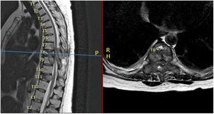
Figure 2.
Sagittal (left) and axial (right) T2-weighted sequence of dorsolumbar MRI showing multiple metastatic lesions with lesion in D8 invading the spinal canal, vertebral body, pedicles, transverse process, and lamina.
Emergency surgical treatment was performed by unipedicular left kyphoplasty D7, D8, and D9 (because the right pedicle was destroyed by tumour mass) and bilateral pedicular D10–D11 (Fig. 3). Minimally invasive “over the top” D8–D9 laminectomy, entering on the right side and performing bilateral decompression through a unilateral approach, with resection of the posterior epidural tumour, of fibrous consistency, highly vascularised, and adherent to the dura mater. Intraoperative blood loss was 40cc and surgical time was 3h.
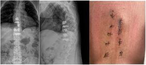
Figure 3.
Postoperative AP (left)/lateral (centre) dorsolumbar X-ray and dorsolumbar percutaneous wounds (right).
The postoperative period was uneventful, and the patient made good general progress over the 10-month follow-up period, with progressive improvement of strength in the lower limbs and recovery of sensory level at the level of D11.
Clinical case 2
A 69-year-old man with a history of stage IV lung carcinoma and overweight, who attended the emergency department for disabling dorsolumbar pain refractory to the usual analgesia. An MRI scan was performed which showed a metastatic L1 lesion without fracture. He was treated by the oncology department with palliative radiotherapy and a dorsolumbar brace. One year after this treatment, he was readmitted for disabling pain without evidence of neurological deficit, with clinical pain, and disability scales VAS 10/10 and ODI 78/100. He was on fentanyl patches 75μg/h and oral morphine sulphate (MST) 90mg/every 12h for pain management.
During his admission, he underwent a further MRI, which showed L1 fracture with posterior wall rupture, invasion of the spinal canal and compression of the conus medullaris (Fig. 4). In addition, there was evidence of multilevel metastatic involvement (D12 involvement with wedging of the superior plate and 2 other foci at L3 and L5, without fracture). He required intravenous perfusion of morphine chloride at 10mg/h for incapacitating pain.
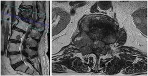
Figure 4.
Sagittal (left image) and axial (right image with cut at L1) T2-weighted sequence of dorsolumbar MRI showing metastatic lesions at D12, L1, L3 with compression of the conus medullaris at L1 level by tumour mass anterior to the thecal sac and by pedicle invasion.
MIS was decided and an “over the top” L1 laminectomy was performed, preferring the right side due to the greater tumour involvement evident on the MRI and bilateral decompression through a unilateral approach, left unipedicular D12 kyphoplasty and percutaneous transpedicular fixation of bilateral D11, left unilateral D12, bilateral L2 and L3 (Fig. 5) with intraoperative blood loss of less than 30cc, and surgery time of 4h.
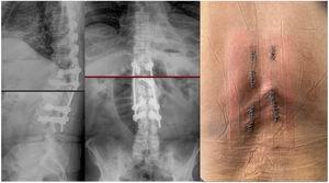
Figure 5.
Postoperative lateral (left image)/AP (centre image) X-rays and percutaneous wounds (right image).
The patient made good postoperative progress, with a follow-up of 6 months, no postoperative complications (neurological deficit or wound dehiscence) and it was possible to progressively decrease intravenous opioids achieving optimal analgesia with oral MST 10mg/every 12h, with postoperative assessment scales of VAS: 3/10 and ODI: 38/100.
Clinical case 3
A 72-year-old woman with a history of ovarian cancer and overweight, admitted to another hospital for disabling dorsolumbar pain managed with usual analgesia and fentanyl patch 50μg/72h, requiring intravenous perfusion of morphine chloride at 6mg/h, and transferred to our centre for treatment. On arrival she presented pain scores VAS: 8/10 and disability (ODI): 68/100. Examination showed no neurological deficit.
Dorsolumbar MRI showed metastatic involvement in the vertebral bodies of L1 and L2; with L2 fracture with subsidence of the right L2 plate. At L1 level, invasion of the spinal canal on the left side and left L1 and L2 pedicle fracture (Fig. 6).
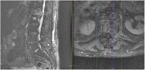
Figure 6.
Sagittal (left image) and axial (centre image with cut at L1) T2-weighted sequence of dorsolumbar MRI showing metastatic lesions at L1 and L2 with compression of the conus medullaris at L1 level by tumour mass anterior to the thecal sac and by left pedicle invasion.
MIS surgery was decided, performing a left L1 hemilaminectomy through a left tubular approach, with a heavily bleeding bone lesion, with difficult haemostasis, right L2 kyphoplasty and elevating the upper plate to improve scoliosis and percutaneous transpedicular cemented fixation of bilateral D11 and D12, right L1 and L2, bilateral L3 (Fig. 7). Intraoperative blood loss of 200cc and surgery time of 4h.
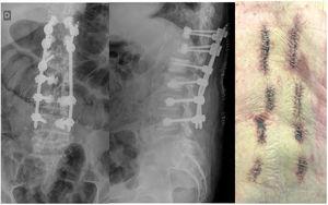
Figure 7.
AP (left image) and lateral (central image) X-rays showing bilateral percutaneous cemented fixation D11–D12, right L1–L2, bilateral L3–L4, and percutaneous wounds (right image).
The patient’s postoperative progress was satisfactory, over a follow-up of 3 months she has not presented postoperative complications, the morphine pump and fentanyl patches have been discontinued. The postoperative clinical scores show improvement of pain VAS: 3/10 and ODI: 20/100.
Discussion
MIS in the treatment of selected cases of spinal metastases has revolutionised the traditional surgical treatment of this disease because it has made it possible to safely and effectively intervene in certain patients who previously might not have been considered surgical candidates. MIS offers advantages over classic techniques, including less blood loss and shorter hospital stays (ICU and general hospitalisation), and a downward trend in complications, while offering similar results to open surgery in terms of pain control (assessed using the VAS scale), overall survival, and improvement or preservation of neurological status (assessed using the ASIA and Frankel scales). Only one study, to date, has reported differences in quality of life in favour of MIS over open surgery, which could infer a greater impact of MIS on the quality of life of patients affected by this devastating disease in the final stages of their lives.21
However, it is important to point out that mastering the MIS technique requires a significant learning curve. This is reflected in the comparative table of MIS vs. open surgery, which does not show a significant difference in surgical time, and MIS involves time, dedication, and training, variables that could interfere with outcomes and prolonged surgery times in the first cases.
In selecting the surgical technique, it is important to emphasise the relevance of the direct and indirect costs of each procedure, so as to determine economic differences when choosing the type of procedure.
Metastatic spinal disease poses an economic challenge for healthcare systems due to the high cost of providing treatment. In Europe, specifically in Denmark, costs range from €36,616 to €87,814 per patient over their survival depending on the treatment received, with specific costs of €36,616 (€33,835–€39,583) for conservative treatment; €49,632 (€42,287–€57,767) for decompression surgery; €70,997 (€62,244–€82,354) for decompression surgery+artrodesis and €87,814 (€76,638–€101,528) for decompression+artrodesis+reconstruction surgery, taking into account that the longer the global survival, the higher the cost of care. It is very important to highlight that hospital stay could represent up to 65% of the total expenditure for the patient, and this item is an objective when it comes to reducing direct costs, followed by follow-up visits, which account for up to 31% of total costs.22
At present, there are no studies available in the databases reviewed that compare the direct costs of MIS vs. open surgery specifically in the treatment of cancer of the spine.23 However, there are studies available comparing the associated costs of both techniques in spinal fusion. Lucio et al. evaluated the difference in hospital costs between MIS and open surgery in spinal fusion procedures in 210 patients, 101 undergoing open surgery and 109 undergoing MIS. They found a higher direct instrumental cost for MIS compared to open surgery at 3810.76US$ (approximately €3500), 27% of the total cost of the procedure. However, they observed that the costs for operating theatres, surgical supplies, and expenses associated with hospital stay was lower for MIS over open surgery by 2756.50US$ (56%), 955.64US$ (45%), and 788.51US$ (52%), approximately €2500, €880, and €726, respectively. They also observed a lower rate of transfusions, re-interventions, and residual events (complications), resulting in an estimated reduction of 2825US$ (approximately €2600) at the end of the hospital and surgical process, and an estimated 10% reduction in the total cost of the surgical process.24
In another study, Vertuani et al. compared spinal fusion in patients undergoing surgery in the UK and in Italy. Their cost analysis included the resources used for each patient and their procedure, including surgical costs (operating rooms, consumables and prostheses, technical equipment, transfusions, surgical drainage), hospital resources (hospital bed and peri-operative medication), and surgical complications. The direct costs derived from surgical material in Italy for MIS was €3137 compared to €2684 for open surgery, with a difference of €453 in favour of open surgery, and in the UK the associated costs were €2856 vs. €2135 with a difference of €721 in favour of open surgery. Despite a trend towards open surgery being more economically favourable in terms of direct costs associated with instruments, in the United Kingdom a lower total cost associated with MIS vs. open surgery was observed (€13,399 vs. €15,065) and the same phenomenon occurred in Italy (€10,012 vs. €10,985), with a difference in favour of MIS of €1666 and €973 for each country, respectively. This is because a decrease in the rate of complications and transfusions (which are more frequent in open surgery), and a reduction in hospital stay compensates for the increase in the unit value of the material for MIS compared to open surgery. However, despite a trend towards cost-effectiveness of MIS, when the value of MIS material exceeds €4111 per patient in Italy and €4578 in the UK, the cost saving benefit for the healthcare system ceases to exist.25 It is vitally important to know the budget required for the optimal treatment of each patient, individualising their requirements, because healthcare costs directly impact the sustainability of the system.
Conclusion
While there is currently no strong evidence supporting the widespread use of MIS for spinal metastases, its benefits in terms of patient recovery, shorter hospital stays, lower infection risk, and reduced postoperative opioid use make it a viable option for selected cases. Prospective randomized clinical trials are needed to further validate its advantages. Additionally, economic studies are lacking but suggest that MIS may lead to lower overall healthcare costs due to reduced complications, transfusions, and shorter hospital stays. However, cost-effectiveness depends on setting limits on instrument costs to maintain the economic benefits of MIS compared to traditional surgery.
BIBLIOGRAFÍA:
[Translated article] The role of Minimally Invasive spine surgery in the treatment of vertebral metastasis (Part 1): A Clinical Review | Revista Española de Cirugía Ortopédica y Traumatología. (s. f.). https://www.elsevier.es/es-revista-revista-espanola-cirugia-ortopedica-traumatologia-129-avance-resumen-translated-article-the-role-minimally-S1888441523001789